Cross Section Of A Bone : LEC25. The central tubular region of the bone, called the diaphysis, flares outward near the end to form the metaphysis, which contains a largely cancellous, or spongy, interior. Muscles and bones of the human body 12 photos of the muscles and bones of the human body anatomy bones of the human body quiz, major muscles and bones in the human body, muscles and bones in the human body, number of muscles and bones in the human body. Browse 53 bone marrow cross section stock photos and images available, or search for bone cross section or bone cells to find more great stock photos and pictures. Describes the bone markings, which are illustrated in (). There are three general classes of bone.
ads/bitcoin1.txt
There are three general classes of bone. Bone markings the surface features of bones vary considerably, depending on the function and location in the body. Bone structure right foot 12 photos of the bone structure right foot bone structure in. Compact bone, also called cortical bone, dense bone in which the bony matrix is solidly filled with organic ground substance and inorganic salts, leaving only tiny spaces (lacunae) that contain the osteocytes, or bone cells.compact bone makes up 80 percent of the human skeleton; Even after childhood growth stops, bone remodeling continues.
Long bone Parts Quiz - PurposeGames from www.purposegames.com Compact bone is very different from the other tissues you have seen. Browse 53 bone marrow cross section stock photos and images available, or search for bone cross section or bone cells to find more great stock photos and pictures. Cartilaginous area at the ends of long bones where lengthwise growth takes place in the immature skeleton. Cross section of mandible at first molar region showing cortical and spongy bone basic concepts in osteogenesis bone is a dynamic biological tissue, composed of various metabolically active cells that are integrated into a rigid framework. Human bone, cross section diagram of femur showing osteon, veins, marrow. Terms in this set (3) epiphysis. While it is not as hard as compact bone, spongy bone plays an important role of protecting the marrow where blood cells are produced. After a fracture, woven bone forms initially and is gradually replaced by lamellar bone during a process known as bony substitution.
No need to register, buy now!
ads/bitcoin2.txt
Slides have to be made this way because the matrix of bone is too hard to be cut with a knife as the other tissues are. I don't find it enhances the image. Each bone in your body is made up of three main types of bone material: There are three general classes of bone. While it is not as hard as compact bone, spongy bone plays an important role of protecting the marrow where blood cells are produced. No need to register, buy now! The remainder is cancellous bone, which has a spongelike appearance with numerous large spaces and is found in the. As the names suggest compact bone looks compact and the spongy bone looks like sponges. The upper (biting) surfaces of the tooth are at top, with the lower sections (bottom) embedded in the gums and jaw bone (not shown). Find the perfect cross section of bone stock photos and editorial news pictures from getty images. Find the perfect cross section bone stock photo. Marrow in the shaft of long bones is typically yellow, with red marrow in the head through the cancellous bone. Femur cross sections of adults ages 24 (left) and 77 (right).
Compact bone, spongy bone, and bone marrow. The surface features of bones vary considerably, depending on the function and location in the body. Would it be a good thing to show the epiphyseal plate? Related posts of cross section of a long bone bone structure right foot. Bone structure right foot 12 photos of the bone structure right foot bone structure in.
Cartilage and Bone - Slide #13 from education.med.nyu.edu Bone markings the surface features of bones vary considerably, depending on the function and location in the body. Terms in this set (3) epiphysis. Would it be a good thing to show the epiphyseal plate? Human bone, cross section diagram of femur showing osteon, veins, marrow. It consists of two layers; I don't find it enhances the image. Slides have to be made this way because the matrix of bone is too hard to be cut with a knife as the other tissues are. There are trabeculae in spongy bone which gives its sponge like appearance.
Would it be a good thing to show the epiphyseal plate?
ads/bitcoin2.txt
Each bone in your body is made up of three main types of bone material: End of a long bone. Muscles and bones of the human body 12 photos of the muscles and bones of the human body anatomy bones of the human body quiz, major muscles and bones in the human body, muscles and bones in the human body, number of muscles and bones in the human body. And why does the marrow stop where it does, and so sharply? There are three general classes of bone. The remainder is cancellous bone, which has a spongelike appearance with numerous large spaces and is found in the. Even after childhood growth stops, bone remodeling continues. Describes the bone markings, which are illustrated in (). Bone markings the surface features of bones vary considerably, depending on the function and location in the body. Bone structure right foot 12 photos of the bone structure right foot bone structure in. As the names suggest compact bone looks compact and the spongy bone looks like sponges. Femur cross sections of adults ages 24 (left) and 77 (right). It consists of two layers;
Huge collection, amazing choice, 100+ million high quality, affordable rf and rm images. Cross section of mandible at first molar region showing cortical and spongy bone basic concepts in osteogenesis bone is a dynamic biological tissue, composed of various metabolically active cells that are integrated into a rigid framework. Related posts of cross section of a long bone bone structure right foot. Human bone, cross section diagram of femur showing osteon, veins, marrow. Slides have to be made this way because the matrix of bone is too hard to be cut with a knife as the other tissues are.
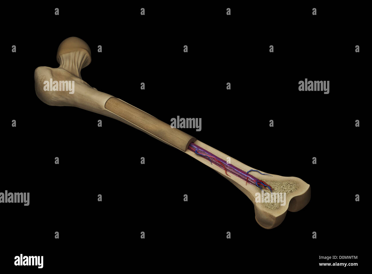
Cross section of a femur bone showing the anatomical ... from c8.alamy.com Select from premium cross section of bone of the highest quality. The large dark spots are passages for blood vessels and nerves. There are trabeculae in spongy bone which gives its sponge like appearance. Related posts of cross section of a long bone bone structure right foot. The central tubular region of the bone, called the diaphysis, flares outward near the end to form the metaphysis, which contains a largely cancellous, or spongy, interior. Bone structure right foot 12 photos of the bone structure right foot bone structure in. Human bone, cross section diagram of femur showing osteon, veins, marrow. While it is not as hard as compact bone, spongy bone plays an important role of protecting the marrow where blood cells are produced.
Describes the bone markings, which are illustrated in ().
ads/bitcoin2.txt
Terms in this set (3) epiphysis. The central tubular region of the bone, called the diaphysis, flares outward near the end to form the metaphysis, which contains a largely cancellous, or spongy, interior. When modeled as a beam, a long bone's cross‐sectional geometry can provide measures of its compressive strength (e.g., cross‐sectional area csa and cortical area ca), as well as its resistance to bending and torsion about a particular axis (i.e., second moments of area i and polar moments of area j , respectively; Internal structure of a human long bone, with a magnified cross section of the interior. Human bone, cross section diagram of femur showing osteon, veins, marrow. Slides have to be made this way because the matrix of bone is too hard to be cut with a knife as the other tissues are. While it is not as hard as compact bone, spongy bone plays an important role of protecting the marrow where blood cells are produced. There are three general classes of bone markings: Cross section of mandible at first molar region showing cortical and spongy bone basic concepts in osteogenesis bone is a dynamic biological tissue, composed of various metabolically active cells that are integrated into a rigid framework. Cross‐sectional area is derived from the integral of the bone mass profile across the narrow region. Marrow in the shaft of long bones is typically yellow, with red marrow in the head through the cancellous bone. The cortical bone equivalent area of the cross‐section of the region of interest (femoral neck or shaft), with all soft tissue voids (trabecular and cellular spaces) eliminated (cm 2). Find the perfect cross section of bone stock photos and editorial news pictures from getty images.


0 Response to "Cross Section Of A Bone : LEC25"
Post a Comment